
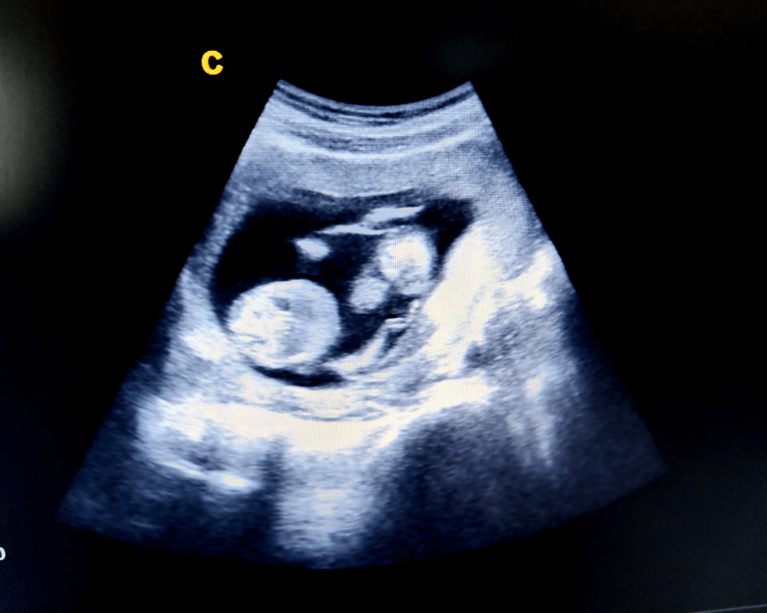
An early structural scan is recommended for all pregnant women in the first trimester of pregnancy.
At Centre for Women’s Ultrasound, we suggest this scan to be performed at 13 weeks to allow for an early detailed look at the baby’s structure and organs.
The early structural scan in first trimester can be extended or combined with:
Nuchal translucency assessment and chromosomal risk assessment
preeclampsia screening for placental function
NIPT for non-invasive fetal chromosome testing
What is a nuchal translucency scan?
The nuchal translucency scan is a non-invasive ultrasound procedure usually performed on the abdomen area, although it may also be performed vaginally. It is performed to measure the amount of fluid present on your baby’s neck and determine if there is any excess fluid. This assessment is important as it allows the diagnosis of potential fetal complications including Down syndrome. If our specialists detect any abnormalities we will notify you of the risks and discuss your options. Counselling and recommendations for non-invasive prenatal testing or definitive tests such as amniocentesis or CVS (invasive confirmation of chromosomal complications) can be offered.
When is it recommended and how is my risk factor calculated?
The nuchal translucency scan is recommended between 12 and 14 weeks of pregnancy as this is the stage of the pregnancy when the thickness of the nuchal translucency fluid layer is seen differently from chromosomally normal fetuses. At The Centre for Women’s Ultrasound we will perform a detailed high quality scan during your first trimester to check the early fetal structure as well as the nuchal translucency and presence of the nasal bone as indicators of a chromosomal issue in the baby.
When determining your risk of chromosomal complications there are three main independent risk factors used in the assessment, including:
- The mother’s age
- The nuchal translucency thickness in the fetus and the presence of the nasal bone
- The mother’s serum level of beta-hCG and PAPP-A
The ultrasound results are combined with a blood test for placental hormones and your age in an algorithm from the Fetal Medicine Foundation, London to perform a risk assessment of a chromosomal anomaly in the baby.
An assessment of fetal heart including tricuspid regurgitance and blood flow in a fetal vessel-ductus venosus is also used in the decision-making.
How accurate is the nuchal translucency scan?
First-trimester Screening program using nuchal translucency scan and blood tests can detect approximately 90% of Down syndrome cases.
As well as Down syndrome detection, a nuchal translucency scan may also detect other abnormalities including:
- Cardiac defects
- Genetic syndromes
- Other chromosomal abnormalities
What is the difference between a nuchal translucency scan and other diagnostic tests?
Non invasive prenatal testing (NIPT) is currently available from 10 weeks for testing of fetal chromosomal abnormalities and is >99% accurate in detecting chromosome anomalies called trisomies such as Down syndrome (Trisomy 21), Trisomy 13 and 18.
The 12-13 week nuchal translucency scan is still recommended as it will check how the baby is forming structurally, as well as pick up other chromosomal anomalies not detected by the NIPT.
Other diagnostic tests are often invasive which can potentially increase your risk of miscarriage (although this increase is considered minimal). A nuchal translucency test can also not provide 100% accuracy, whereas other diagnostic tests, in some cases, can. Our specialists recommend either having a nuchal translucency (non-invasive) scan first, or NIPT followed by a first trimester morphology/nuchal translucency scan. If we believe further diagnosis or confirmation is necessary we may recommend a chorionic villus sampling (CVS) or amniocentesis test.
Our Technology
The Centre for Women’s Ultrasound is a centre for excellence when it comes to performing first trimester Down syndrome screening using Fetal Medicine Foundation software from London, UK.
Do you have a question about nuchal translucency scans? Please contact the Centre for Women’s Ultrasound today.
Common questions
Can't find your answer here? Our team is happy to help before your appointment.
The Nuchal Translucency (NT) measurement, obtained during the Nuchal Translucency scan between the 11th and 14th weeks of pregnancy, indicates the thickness of the clear space at the back of the baby’s neck. This measurement is instrumental in assessing the risk of chromosomal abnormalities, particularly Down syndrome, as well as potential cardiac anomalies. An increased NT measurement, in conjunction with maternal age and other factors, may suggest a higher likelihood of such conditions. However, it is crucial to emphasise that the NT measurement is part of a screening process, not a definitive diagnosis. Further diagnostic tests may be recommended for confirmation, and counselling is provided to parents to navigate their options and understand the broader context of prenatal care.
Several factors can influence the accuracy of the Nuchal Translucency (NT) measurement during pregnancy, including the gestational age at which the scan is conducted (ideally between the 11th and 14th weeks), the baby’s position during the scan, maternal characteristics such as obesity, the complexity of twin pregnancies and individual foetal positions, the presence of foetal abnormalities affecting the neck or head region, the precision of the Crown-Rump Length (CRL) measurement, and the experience of the ultrasound operator. These variables underscore the importance of careful consideration and communication by healthcare providers to manage expectations and guide decision-making regarding the interpretation of NT measurements and potential follow-up diagnostic tests.
After the Nuchal Translucency (NT) scan, you can expect to receive information about the measurements taken during the scan, particularly the thickness of the clear space at the back of the baby’s neck. The healthcare provider will typically combine the NT measurement with other factors, such as maternal age and blood test results, to estimate the risk of chromosomal abnormalities like Down syndrome. It’s important to note that the NT scan is a screening test, not a diagnostic one. If the results indicate an increased risk, your healthcare provider will discuss the findings with you, provide counselling, and offer options for further diagnostic tests, such as chorionic villus sampling (CVS) or amniocentesis, which can provide more conclusive information about the baby’s health. The timing of receiving results can vary, but healthcare providers often aim to communicate findings promptly to allow for informed decision-making and further planning. It’s crucial to discuss specific timelines with your healthcare provider during or after the NT scan.